
Vastus medialis linea aspera of femur (origin) medial quadriceps tendon to patella, tibial
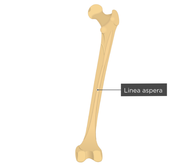
The linea aspera is the rough, longitudinal crest on the posterior surface of the femoral shaft. Most orthopedic surgeons depend on the linea aspera as an intraoperative landmark identifying the true posterior aspect of the femur. We investigated the position of the linea aspera to verify whether the surgeon can rely on this accepted belief.

Linea Aspera Anatomy Anatomical Charts & Posters
Calcification at the linea aspera is a differential for hip/thigh pain. • Imaging should commence with plain radiographs, and also include the distal femur. • Smaller calcific deposits can be obscured on anteroposterior projection, necessitating additional views. • Calcific tendo-enthesopathy is associated with osteolysis but no soft.

Calcification of the linea aspera A systematic narrative review European Journal of Radiology
The linea aspera (Latin: rough line) is a ridge of roughened surface on the posterior surface of the shaft of the femur. It is the site of attachments of muscles and the intermuscular septum.. Its margins diverge above and below. The linea aspera is a prominent longitudinal ridge or crest, on the middle third of the bone, presenting a medial and a lateral lip, and a narrow rough, intermediate.
Femur Linea Aspera Anatomy Tibia Intertrochanteric Line, PNG, 853x886px, Watercolor, Cartoon
Plain radiograph. On anteroposterior projections of the femur in adults and rarely, in adolescents, the linea aspera may appear as two axially-oriented, parallel lines in the middle of the femoral shaft. This appearance, termed the track sign, is a normal variant that is important to distinguish from the blade of grass sign in Paget disease 2.

Vastus intermedius (O) anterior surface of femur & linea aspera (Ins) patella and tibial
The linea aspera, also referred to as the linea aspera-pilaster complex, is a characteristic ridge that runs along the posterior aspect of the human femur. While much less pronounced in the non-human primate than in the adult human femur (Hrdlička 1934 ), the linea aspera-pilaster complex imparts a unique triangular "peak" to an.
Linea aspera hires stock photography and images Alamy
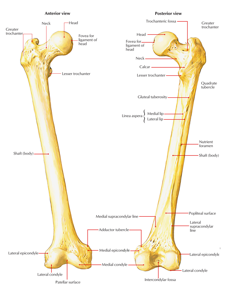
Linea aspera, located on the posterior border of the femur's body or shaft, is a rough bony ridge. It serves as an attachment site for intermuscular septa and various muscles. The adductor magnus, the largest adductor muscle, inserts directly onto the linea aspera and its extensions above and below. The linea aspera features prominent medial and lateral lips, which offer origin to the vastus.
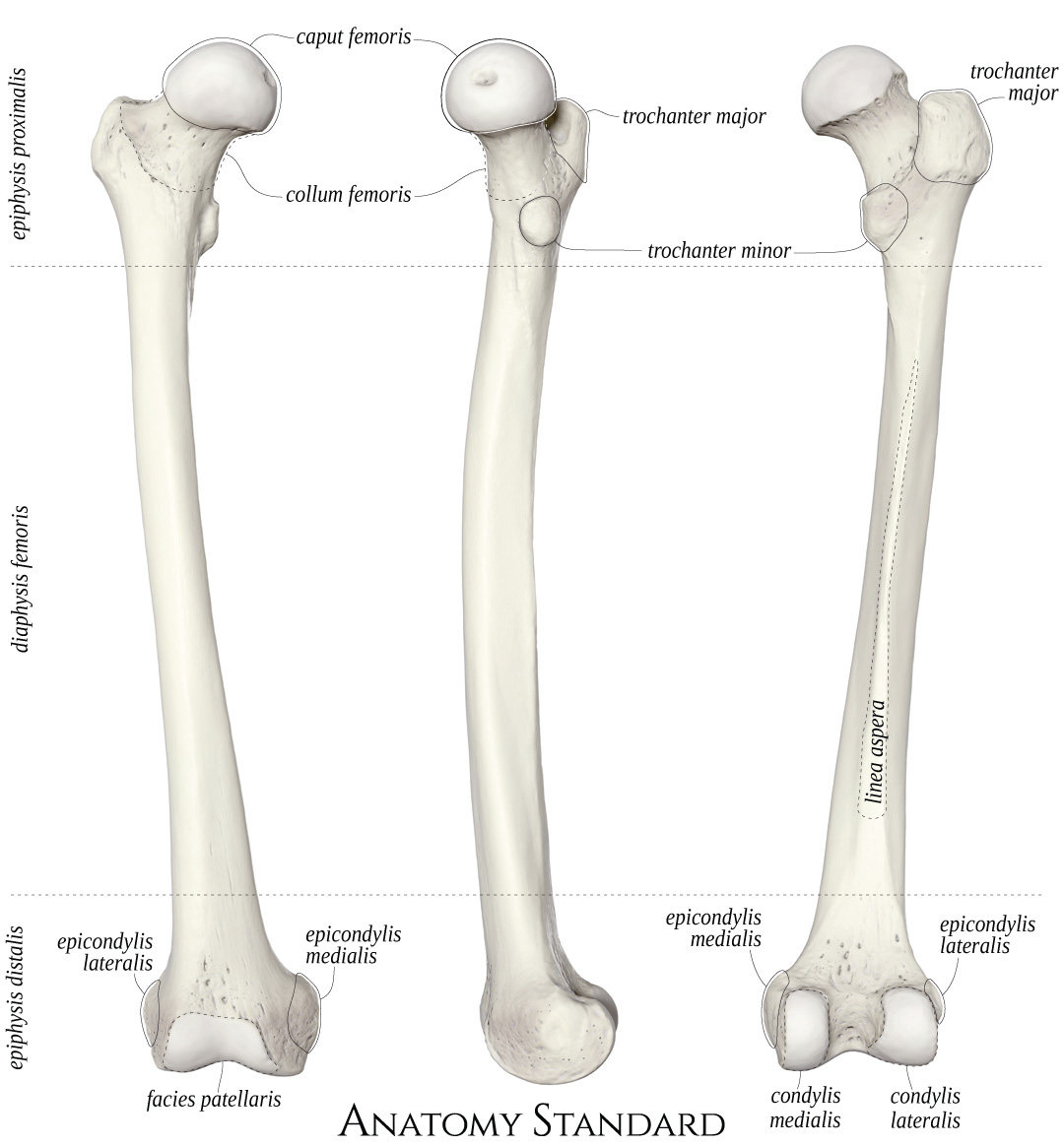
Anatomy Standard Drawing Femur anterior, medial and posterior view Latin labels AnatomyTOOL
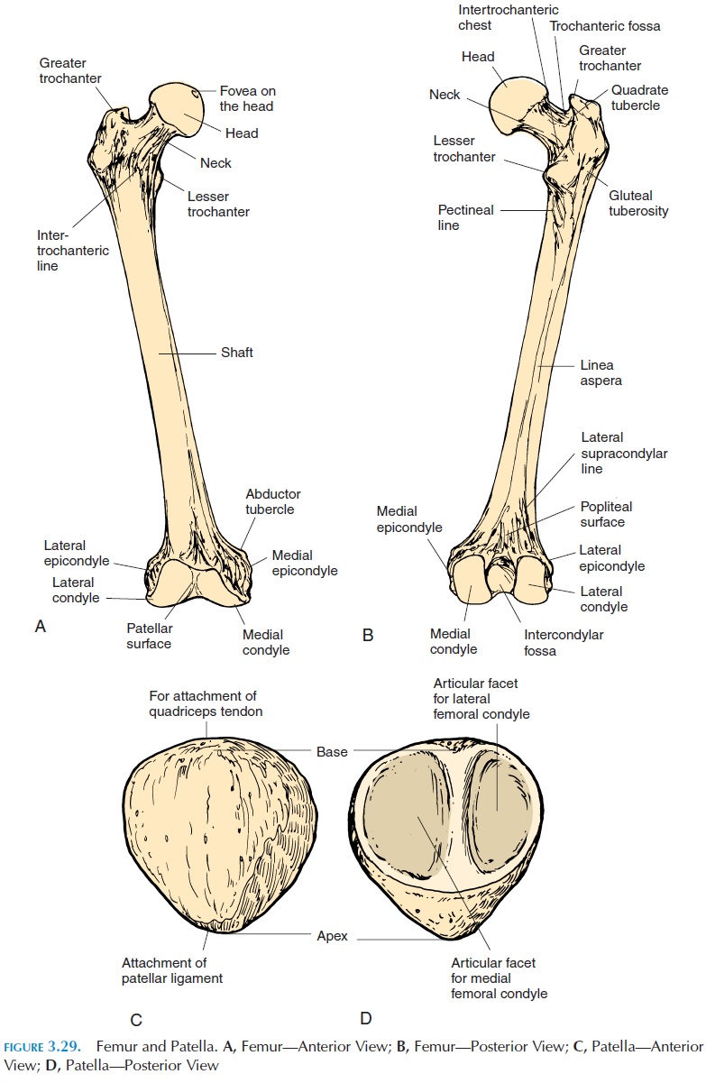
The groove is continuous with the lateral lip of the linea aspera. On the medial, proximal, posterior part of the femur is another (smaller ridge) known as the pectineal line. It acts as the point of attachment for the pectineus muscle. The most pronounced part of the posterior surface is the linea aspera. This is a raised longitudinal.
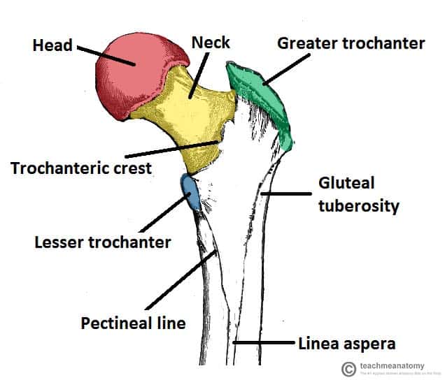
The Femur Proximal Distal Shaft TeachMeAnatomy
At the extremity of the medial supracondylar line, there is an adductor tubercle. The medial border of the linea aspera meets the pectineal line, whereas its lateral border gives rise to the gluteal tuberosity. Towards the distal end, the linea aspera widens and forms the floor of the popliteal fossa. 3. Distal Femur. The distal end of the.

Learn Femur diagram (by wawezase) Remember and Understand
Gluteal tuberosity is the upper lateral continuation of the linea aspera as it extends towards the greater trochanter of the femur. It presents as a roughened bony ridge that serves as the site of attachment for the gluteus maximus muscle.

The Quadriceps Muscles Yoganatomy
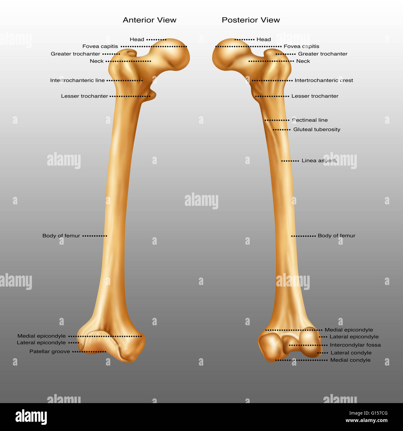
On the posterior surface of the femoral shaft, there are roughened ridges of bone, called the linea aspera (Latin for rough line). This splits distally to form the medial and lateral supracondylar lines. The flat popliteal surface lies between them. Proximally, the medial border of the linea aspera becomes the pectineal line.
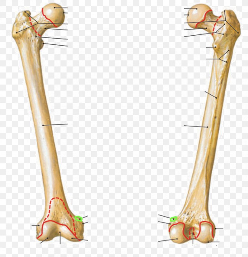
Femur Bone Posterior Markings
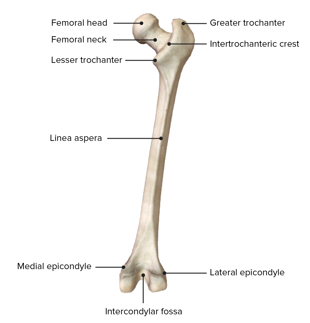
The linea aspera ( Latin: rough line) is a ridge of roughened surface on the posterior surface of the shaft of the femur. It is the site of attachments of muscles and the intermuscular septum. Linea aspera. Right femur. Posterior surface. (Linea aspera not labeled, but region is visible. Medial lip is at left; lateral lip is at right.)

SAR HS 369 Lecture Notes Summer , Lecture 12 Intertrochanteric Crest, Lesser Trochanter
Pectineus muscle inserts into the posterior surface of femur, along the pectineal line and proximal part of linea aspera. These two aforementioned lines are continuous with each other; the pectineal line continues inferiorly from the intertrochanteric line and ends by fusing with the spiral line of femur, thus forming the medial lip of linea.

Femur Thumb Structure Bone Linea Aspera PNG, Clipart, Abdomen, Angle, Arm, Bone, Elbow Free PNG
The linea aspera (LA) is the distinctive ridge found along the posterior aspect of the femur. When translated from Latin, LA means "rough line.". LA is the roughened, longitudinal irregular crest that is composed of 2 lips. This feature is the insertion site of the adductor thigh muscle. It is the origin of several muscles in the thigh. [2.
Femur Earth's Lab
The borders of the femur are the linea aspera, a medial border, and a lateral border. Linea aspera border. The linea aspera is a prominent longitudinal ridge or crest, on the middle third of the bone, presenting a medial and a lateral lip, and a narrow rough, intermediate line. Above, the linea aspera is prolonged by three ridges.
Thigh Anatomy Concise Medical Knowledge
The linea aspera is made up of a medial lip, a lateral lip, and an intermediate line. The linea aspera diverges at the proximal and distal ends of the posterior femoral body. Proximally, the lateral border of the linea aspera becomes the gluteal tuberosity (mentioned above), and the intermediate line becomes the pectineal line (mentioned above).
Linea Aspera Femur
Then, each femur was aligned to its anatomical axis before the femoral anteversion and linea aspera version were measured. Images were displayed in axial, sagittal and coronal views. Image of the femur was isolated from the surrounding soft tissue and other bone parts using thresholding, region growing, and segmentation tools available in the.